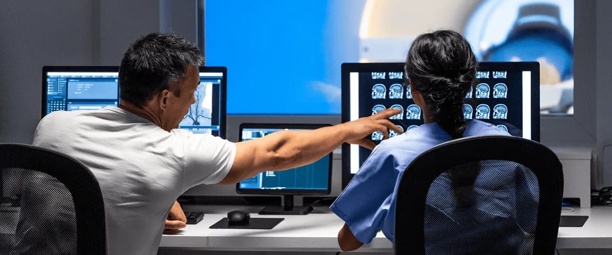
Magnetic resonance imaging (MRI) has revolutionised the field of neuroscience, providing high-quality images of the brain and spinal cord. Neurological disorders are much easier to diagnose with this tool. In this article, we highlight the principles, advances and applications of MRI in neurology for the benefit of specialists in this medical niche.
Magnetic resonance imaging technology has evolved in recent years, making it possible to visualise the structure and function of the nervous system in detail. Serious neurological diseases such as epilepsy, Alzheimer’s, brain tumors or multiple sclerosis can be diagnosed quickly and treated more effectively thanks to MRI.
What is MRI
Magnetic resonance imaging is an advanced non-invasive imaging technology that uses a magnetic field and radio waves to produce detailed, three-dimensional anatomical images. It is widely used across medical fields such as neurology, cardiology, oncology, orthopaedics, gastroenterology, obstetrics-gynaecology and other specialties. Unlike CT, X-rays or ultrasound, MRI produces clearer images of tissues, fat or body fluids.
Types of neurological MRI
There are two main types of neurological MRI:
- Diffusion-weighted imaging (DWI) MRI used to detect acute strokes where brain tissue begins to deteriorate or die due to lack of blood and oxygen.
- MRI spectroscopy (MRS) used in certain neurological conditions (multiple sclerosis or tumors) to detect and analyse chemicals in the brain.
The MRI typology also shows that there is contrast MRI which involves injecting a contrast agent into the blood to make certain structures more visible as well as functional MRI which measures brain activity by detecting changes in blood flow during certain cognitive and behavioural activities.
Depending on the neurological area being examined, there is brain MRI which visualises blood vessels in the head, middle and inner ear and anatomical components as well as spinal MRI which provides precise information about intervertebral discs, ligament and vertebral integrity, spinal nerves, etc.
Applications of MRI in neurology
An MRI produces detailed images of multiple structures: cranial nerves, posterior fossa abnormalities, spinal cord, brainstem lesions. For this reason, it is essential for neurological diagnosis, treatment planning and monitoring of central nervous system disorders.
Magnetic resonance imaging provides high-resolution morphological images, unique functional information and superior contrast of soft tissues in the central nervous system. MRI is superior to other imaging modalities used in neurology for detecting spinal abnormalities (tumors, abscesses), demyelinating plaques, subclinical cerebral oedema, early infarction, cranio-cervical junction abnormalities, cerebral contusions, syringomyelia or trans-tentorial herniation.
The excellent contrast between grey matter and white matter makes the examination a very good choice for various central nervous system conditions such as dementia, demyelinating diseases, cerebrovascular diseases, epilepsy and infectious diseases. It can also be used to study functional and structural abnormalities in psychological disorders.
MRI is also a very useful investigative tool in neurological cancers offering a better resolution than CT and superior visualisation of the posterior fossa. It is also widely used in stereotactic MRI-guided surgery and radiosurgery, in the treatment of intracranial tumors or arteriovenous malformations.
MRI-detected neurological disorders
A neurological MRI can detect many health problems of the brain and central nervous system including:
- Stroke & cerebral ischemia
- Multiple sclerosis
- Brain tumors
- Epilepsy
- Blood vessel diseases
- Traumatic brain injury
- Dementia
- Congenital anomalies
- Brain & spinal cord infections
- Parkinson’s disease
- Spinal cord & spinal column disorders
- Vascular malformations
When would doctors recommend MRI?
Neurologists and healthcare providers may recommend an MRI of the brain in several situations, both to diagnose certain conditions and to monitor existing conditions.
Certain patient symptoms may prompt specialists to request an MRI: These include chronic migraines or headaches, frequent episodes of dizziness, vision problems that cannot be explained by eye exams, hearing loss without an explainable cause, radical changes in behaviour and thinking, hormonal imbalances related to the pituitary gland or hypothalamus, weakness and extreme fatigue.
In addition to these, healthcare providers also indicate MRI scans before brain surgery and after surgery to make sure the healing process is going well.
Who performs a brain MRI
A brain MRI can be performed by a radiologist or radiology technologist. The radiologist specialises in performing and interpreting imaging tests. The technologist is a healthcare provider who has been trained to perform such a test and is certified to perform MRI scans. The actual scan takes about 30 to 60 minutes, and in the case of contrast MRI the time may be extended.
How an MRI brain scan works
The MRI technique involves passing an electric current through coiled wires that create a temporary magnetic field in the body. The transmitter and receiver send and receive radio waves, and a special computer uses these signals to create images of brain structures.
What are the latest findings in neurological MRI?
Recent MRI findings have been very useful for healthcare professionals. For example, the use of functional MRI has shown that the human brain has an amazing ability to re-organise and adapt, allowing rapid recovery from disease and injury. At the same time, MRI has enabled researchers to map the connections between brain regions more precisely.
Using MRI at the molecular level allows researchers to more accurately identify biochemical markers of brain disease such as amyloid in Alzheimer’s disease.
MRI-based neuroimaging techniques have led to the discovery of modern treatments for certain conditions such as transcranial magnetic stimulation therapy for depression, anxiety and panic attacks.
Conclusion
MRI technology is particularly useful for healthcare professionals in diagnosing neurological conditions, monitoring their progression and checking the effectiveness of treatments.
Today there have been huge advancements in image resolution which help in the early detection of many conditions, including brain tumors, as well as reducing the time it takes to perform the procedure. MRI is an essential tool that is constantly being improved but research and development in this area is desirable in the future to further help patients with neurological conditions.


