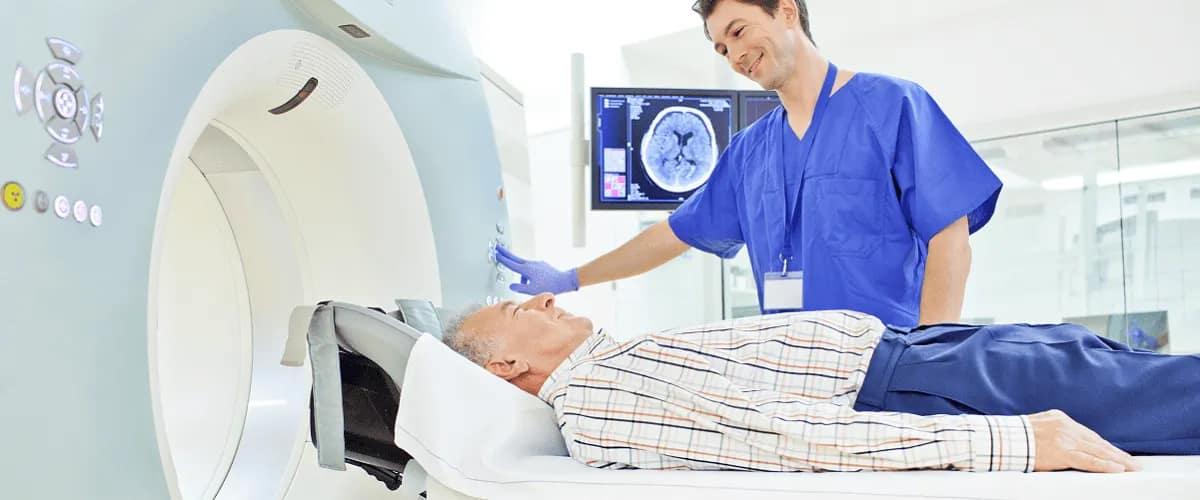
Computed tomography (CT) is one of the cutting-edge medical technologies that has revolutionised the way doctors diagnose and treat cancer. This article details the benefits of using this method of medical investigation. Early detection of tumours and increased life expectancy for patients diagnosed with cancer are among the issues addressed.
Early detection of cancer is crucial to successful treatment and reducing the risk of complications. A CT scan can accurately detect changes in the structure, shape and size of tissue or tumours long before the patient experiences symptoms. This allows the clinician to develop an immediate treatment plan that will increase the patient’s life expectancy.
What does CT mean?
Computed tomography (short: CT) is a medical imaging method that uses X-rays to produce detailed images of organs, tissues and internal structures of the human body. These images are highly accurate and are used to diagnose and treat various medical conditions such as cancer, cardiovascular disease, bone disease and traumatic brain injury.
How does the CT examination work?
Technically, it is carried out using a cylindrical device inside which X-rays are generated around the patient whose body absorbs the radiation beams. An X-ray detector measures the intensity of X-rays reaching the tissues and converts them into an electrical signal. The signal is then processed by a computer to generate a detailed image of the body section being scanned.
CT: An invaluable aid in detecting cancerous tumours
Cancer is one of the most dangerous diseases facing people today. In such situations the clinician has an enormous responsibility in determining the treatment plan. However, this cannot be done without an accurate diagnosis which often only a CT scan can guarantee.
CT is able to identify tumours, abnormal changes in organ size and other problems that may be signs of cancer. One of the biggest advantages of CT scans is that they can detect tumours that are too small to be seen by physical examination or other types of scans such as X-rays or ultrasound. By identifying these tumours at an early stage doctors can intervene earlier and treat patients before the tumour spreads and becomes more difficult to treat.
Which areas of the body can be investigated with CT?
Computed tomography can be used in various areas of the body such as the brain, lungs, liver, kidneys, organs of the reproductive system and parts of the bone system. The area of cancer diagnosis therefore covers malignant tumours located in almost all regions of the human body.
CT-guided biopsy
Accuracy is the key word that defines CT technology. After a CT scan the tumour can be localised to the millimetre in a very short time. This allows a biopsy to be performed quickly, where a sample of suspicious tissue is taken and examined in the laboratory to determine its nature. The CT scan can be used to guide the biopsy allowing the doctor to take a sample that is relevant to the diagnosis.
Checking the results of cancer treatment by CT
Whatever treatment is prescribed—be it chemotherapy, radiotherapy, surgical removal of the tumour, immunotherapy or cancer-specific therapies—it is important to check the effectiveness of the treatment regularly.
CT can be used to monitor a patient’s response to cancer treatment. CT scans can show whether the tumour has shrunk or whether there is a positive response to treatment. Depending on the progress of the tumour, the doctor will then decide whether a change in treatment is needed or whether treatment should continue.
Precise surgical removal of the tumour using CT
Computed tomography can be used during surgery to guide the surgeon during the procedure. CT images can help the surgeon see the exact position of the tumour and decide the best way to remove it. This is particularly useful if the tumour is located in a sensitive or difficult-to-access area such as the brain or organs and tissues in the chest cavity.
Targeting radiotherapy
Computed tomography also plays an important role in the treatment of cancer by radiotherapy. It allows the doctor to visualise the tumour and determine its exact position in the body, so that the radiotherapy can be precisely targeted to the affected area. This reduces the risk of irradiating other healthy tissues and organs around the tumour and minimises unwanted side effects.
Advantages of CT
As with any medical procedure, CT has advantages and disadvantages that must be taken into account when making decisions about its use and recommendations for patients. These include:
- The ability to produce detailed and highly accurate images of internal body structures, bones, soft tissues and internal organs. In this respect, it is a highly accurate imaging technique superior to other examination technologies.
- Depending on the area examined, the investigation can be completed in up to 30 minutes which is extremely useful for patients who have difficulty sitting still for a longer period of time.
- Non-invasiveness: CT examination does not require incisions or anaesthesia—procedures that could lead to further complications or recovery time. If the doctor recommends the administration of a contrast agent, by injection or swallowing, it will be easily removed afterwards.
Disadvantages of CT
Disadvantages of using CT may relate to the following aspects:
- Allergic reactions to the contrast agent. These are very rare and range from mild skin reactions to severe reactions. Reviewing the patient’s medical history before the examination reduces the risk of allergic reactions.
- Toxicity of the contrast agent to the patient’s kidneys, if the patient suffers from renal failure.
- Radiation exposure: However, experts say that the doses of radiation used in CT scans are relatively low and cannot be considered a significant risk for developing cancer later.
All in all, CT is an important imaging method in the diagnosis and treatment of various types of cancer. This technology provides detailed images of internal organs and tissues allowing doctors to detect the presence of tumours, determine their size and location and assess the degree of malignancy.


