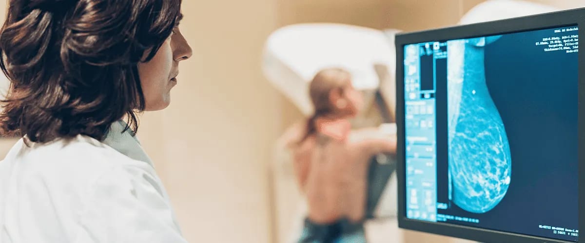
Mammography is a vital tool for the early detection of breast cancer which is the most common cancer in women worldwide and a serious public health concern. Medical professionals need to understand this method of investigation as it plays an essential role in the prevention, screening and detection of cancer.
For successful treatment, it is important that breast cancer is detected at an early stage. With a thorough understanding of the benefits and limitations of this screening method, healthcare professionals can make informed decisions with the patient.
What is a mammogram?
A mammogram is an X-ray investigation of the breasts that is crucial in detecting cancer in women who may or may not have symptoms of a breast problem. It detects the disease at an early stage, before lumps and cysts can be felt by palpation.
Mammograms may be recommended for screening, i.e. as a routine check to detect changes (such as calcifications, distortions, masses, etc.) and see if there is any cause for concern. If the patient has symptoms specific to breast cancer, mammography becomes a diagnostic tool.
Although it uses X-rays, it is considered a safe method of investigation, as the patient is exposed to a negligible amount of radiation during the examination. The procedure can cause some discomfort as the breasts are compressed.
The degree of discomfort depends on the size of the breasts, the skill of the technologist and the sensitivity of the breasts (breasts may be more sensitive during the premenstrual or menstrual period).
When is mammography recommended?
In general, medical experts believe that women should have mammograms from the age of 40 every one or two years, depending on the doctor’s advice. The timing varies depending on the risk of developing breast cancer in women with a family history of breast cancer.
Along with breast self-examination, mammography plays a crucial role in the early detection of breast cancer. The decision about how often to have a mammogram is made with the doctor. Risk factors and individual circumstances will be taken into account.
Early detection means a better prognosis, as treatment is more likely to be successful in the early stages of cancer. Early detection also means that the cancer is less likely to spread to other parts of the body.
The mammography images can also help the doctor to determine whether the patient is likely to develop breast cancer in the future.
How are mammogram results interpreted?
BI-RADS is a standard system used to describe the results of a mammogram. This score indicates the degree of malignancy and whether further tests are needed. For example, a BI-RADS score of 0 means that the results are inconclusive and the radiologist recommends a new examination.
In this case, mammography may be repeated or other imaging tests, such as ultrasound, may be recommended. A score of 1 is a normal score and indicates that the images show no abnormality in the breast tissue.
At the other end of the scale, a score of 5 means that the abnormalities found are very malignant and a biopsy will be taken. A score of 6 is given if a cancerous tumour has already been detected in a previous biopsy and mammography is carried out to monitor the disease after treatment.
If patients have a normal result, it is important that they continue to follow their doctor’s recommendations about how often they should repeat the mammogram.
If the mammogram shows an abnormality, it does not necessarily mean that the patient has breast cancer. Only follow-up tests can confirm whether it is cancer or not.
Mammography risks & limitations
Although mammography is an important imaging procedure in the early detection of breast cancer, it bears some risks and limitations. The first is radiation exposure, but as described above the dose is very low.
In addition, abnormalities are more difficult to see in patients with breast implants, and full visualisation of the breast tissue may not be possible. This is where the skill of the technologist is particularly important as they need to know how to perform the examination in people with breast implants.
One limitation of mammography is that it can give false negative or false positive results. These situations are rare, but it is good for healthcare professionals to be aware of this discrepancy.
Also, if the cancer is located in an area that is difficult to visualise accurately during the mammogram, it may be missed in the interpretation.
Mammography procedure
Before the procedure, patients will be asked to remove their clothing from the waist up as well as their jewellery. They will wear a gown and lie or stand. The breast is placed on a platform and then flattened by the action of a plate designed to compress the breast and flatten the tissue.
This is the only way to get a clear picture of the breast tissue. The same procedure is repeated for each breast and the compression may be repeated several times to obtain images of the sides of the breast.
To ensure that the images are clear, the technologist may ask the patient to hold her breath while the images are taken. The patient should also not wear deodorant, perfume or other products that may affect the clarity of the images and show up as white spots.
After completing the procedure, the technologist will review the images to ensure they are sufficiently clear. If not, additional images will need to be taken. The entire procedure takes approximately 20 minutes, and the potential discomfort is short-lived when breast compression is performed.
All in all, mammography is an important tool in the early detection of breast cancer. It is vital to understand the benefits and limitations of this X-ray procedure in order to provide a good quality of care for patients and ensure that more women benefit from early detection.


