Positron emission tomography (PET) imaging, used primarily in cancer screening but also in cardiology and neurology, has recently demonstrated its effectiveness in the diagnosis and treatment of Parkinson—a neurodegenerative disorder that affects an estimated 11 million people worldwide.
Parkinson is a progressive disease that affects the central nervous system and causes symptoms such as tremors, muscle stiffness, postural instability and problems with voluntary movements. This article discusses the application and benefits of PET in the clinical management of the Parkinson.
A PET scan provides an accurate diagnosis of Parkinson’s disease—eliminating the possibility of confusing symptoms with other neurodegenerative disorders, identifying the affected area of the brain, and developing an appropriate treatment plan.
What is PET imaging?
PET or positron emission tomography uses radioactive substances to visualise the blood flow and functioning of organs and tissues in the human body. This process is made possible because radioactive substances injected into the patient’s veins emit positrons that are detected by the PET scanner.
The method is used to diagnose serious diseases such as cancer, heart disease and neurological disorders. In the case of Parkinson’s disease, a PET investigation can be very useful in determining the correct diagnosis and choosing the right treatment.
PET procedure in Parkinson patients
- During the PET procedure, a radioactive substance called tracer is injected into the patient’s bloodstream, where it is taken up by the circulatory system and transported to the brain and other parts of the body.
- The scanner produces a 3D image of the brain showing the distribution of the tracer in different areas. Depending on how the substance spreads and behaves, the pathology the patient is suffering from can be determined more easily.
- There are several PET tracers available that can be used to understand how Parkinson’s disease works, its stage, the degree of motor impairment, progression, response to treatment, and metabolic changes in different areas of the brain.
- An imaging agent called 18F-DOPA PET can be used in patients with motor symptoms associated with Parkinson’s disease. The substance is a precursor of dopamine, a neurotransmitter or cell signalling molecule that is severely reduced in Parkinson patients.
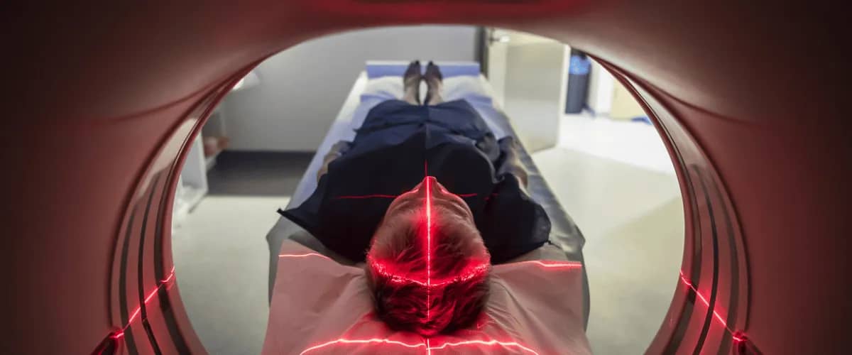
- This is due to the death of dopamine-producing nerve cells in a region of the brain called the substantia nigra. The scan is carried out in the same medical device used for a CT scan, i.e. in a cylindrical tube in which the patient lies horizontally.
- The computer generates 3D images of the brain that can be viewed on a monitor and show how the tracer is distributed and acts in different parts of the brain.
- Based on specific patterns of 18F-DOPA uptake in the dark matter region of the brain, the doctor can determine the presence and stage of the disease as well as the appropriate treatment.
PET to distinguish neurodegenerative diseases
One of the most important benefits of PET in the management of Parkinson’s disease is that it can help differentiate between Parkinson and other neurodegenerative diseases.
Parkinson symptoms can often be confused with those of other conditions such as essential tremor, dystonia or atypical parkinsonian syndrome. A medical PET scan is used to identify subtle differences in brain structure and function that may indicate the presence of Parkinson’s disease.
Pinpointing the area of the brain affected by Parkinson
Another major benefit of PET is the ability to pinpoint the exact area of the brain affected by Parkinson. Depending on its size and location, the doctor can choose the best treatment. For example, in patients with severe Parkinson symptoms, the doctor may decide to recommend deep brain stimulation.
This involves implanting a device in the brain to help reduce the symptoms of the disease. The device is implanted surgically, a very delicate operation with success depending on precise knowledge of the area where intervention is needed to correct the movements affected by Parkinson’s disease.
Medical PET scanning can help the doctor to identify this area more precisely and minimise the risk of complications.
PET: A reliable tool in Parkinson treatment
Medical investigation can be successfully used to monitor the progress of the disease and assess the effectiveness of the treatment prescribed by the neurologist.
PET is used to estimate the level of dopamine in the brain, a neurotransmitter that plays an important role in the control of voluntary movements. In Parkinson’s disease, dopamine production is impaired, which can lead to the development of symptoms specific to the disease.
PET scans can be used to assess dopamine levels in the brain and help identify areas affected by the disease. This can be very useful in choosing the appropriate treatment and monitoring the progression of the disease over time.
Sometimes, the prescribed treatment may not have the effects expected by the doctor—proving to be ineffective or even harmful because of side effects caused by inadequate dosage or the patient’s intolerance to the substances administered.
For example, one of the treatments used for Parkinson’s disease involves taking L-dopa or levodopa, a precursor of dopamine that can help reduce the symptoms of the disease.
However, treatment with this drug can lead to unwanted side effects such as dyskinesia, which is an abnormal movement of the body. The PET scan can help the doctor identify the areas of the brain that are affected by dyskinesia and adjust treatment accordingly.
Early Parkinson detection
PET scans can also be used to identify patients who are at higher risk of developing Parkinson’s disease. For example, people who have a family history of Parkinson’s disease or who have a particular genetic mutation may be more likely to develop the condition.
The use of medical PET investigation can help identify these individuals and intervene at an early stage of the disease, which can lead to an improved prognosis and quality of life.
All in all, PET is a particularly useful medical investigation in the diagnosis and treatment of Parkinson’s disease—helping to differentiate the symptoms of this disease from those of other neurodegenerative diseases, to precisely identify the affected areas of the brain, and to monitor the progress of the treatment.
Therefore, PET is a valuable tool to help physicians make informed treatment decisions and improve the quality of life for patients diagnosed with Parkinson’s disease.


