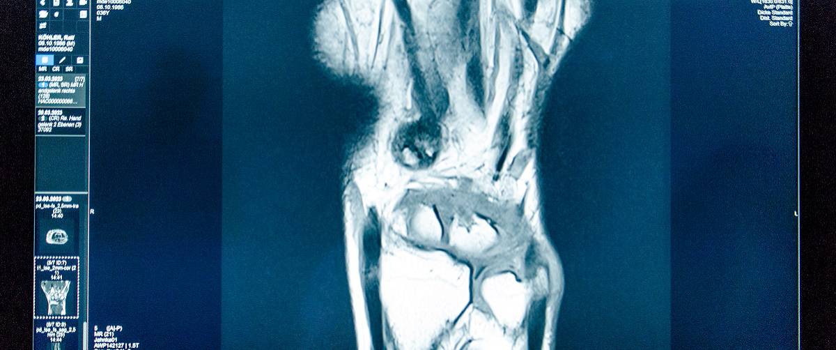
A DICOM viewer is a computer application designed to access, display, and manipulate medical images stored in the DICOM format. Learn more about it and where it is used.
📖 Author: Emmanuel Anyanwu | Alberta Health Services, Canada
In healthcare, medical imaging plays a pivotal role in diagnosis, treatment planning, and monitoring patients’ progress.
With the advent of digital technology, the need for efficient and standardised image management led to the development of DICOM (Digital Imaging & Communications in Medicine) viewers, also known as PACS (Picture Archiving & Communication System) viewers.
A DICOM viewer is a computer application designed to access, display, and manipulate medical images stored in the DICOM format. DICOM is an internationally recognised standard for storing and exchanging medical images and related information.
DICOM viewers facilitate the interpretation of various medical images, including X-rays, CT scans, MRI scans, ultrasounds, and more. These software tools enable healthcare professionals, like radiologists, physicians, and clinicians, to visualise and analyse images on computer screens.
Functionality of DICOM viewers
DICOM viewers provide a range of powerful features and functionalities that aid in medical image interpretation. Here are some essential functions commonly found in DICOM viewers:
- Image viewing: DICOM viewers allow users to navigate through medical images, zoom in and out, pan across the image, and adjust parameters like brightness and contrast. These tools enhance the visualisation of anatomical structures and help identify abnormalities.
- Measurement & annotation: DICOM viewers enable users to directly measure the images’ distances, angles, and other quantitative aspects. Additionally, annotations can be added to highlight specific areas, annotate findings, or communicate observations effectively.
- Multi-planar reconstruction (MPR): DICOM viewers offer MPR capabilities, allowing users to reconstruct images in different planes (axial, coronal, sagittal) for a comprehensive examination. This feature aids in understanding complex anatomical structures and identifying abnormalities from multiple perspectives.
- 3D volume rendering: Advanced DICOM viewers support 3D volume rendering, which allows the creation of three-dimensional representations of anatomical structures. This feature enhances the visualisation of intricate structures—aiding in surgical planning and preoperative assessments.
- Integration with PACS systems: DICOM viewers often integrate with PACS systems which are the central repositories for medical images and associated patient data. The integration allows users to access patient studies, retrieve images, and synchronise data seamlessly—eliminating the need for manual transfers between systems.
Applications of DICOM viewers
DICOM viewers find extensive applications in healthcare facilities where medical imaging is performed. These include:
- Radiology departments: DICOM viewers are used all over radiology departments to view and assess images from different modalities and in PACS. Radiologists use DICOM viewers to interpret and analyse various medical images, providing accurate and timely diagnoses.
- Clinical practices: Physicians and clinicians utilise DICOM viewers to review medical images and collaborate with radiologists for comprehensive patient assessments.
- Medical imaging centres: DICOM viewers are essential tools in imaging centres—aiding in the visualisation and interpretation of images acquired from various modalities.
- Teaching & research: DICOM viewers facilitate educational activities—enabling instructors and researchers to study and share medical images for academic purposes.
Installing a DICOM viewer
Installing an independent DICOM viewer depends on the specific software and platform. Here is a general overview of the installation process:
- Identify a DICOM viewer that suits your requirements and check its compatibility with your operating system.
- Contact vendor or visit the vendor’s website or a trusted software repository to acquire the DICOM viewer software.
- Follow the provided instructions to initiate the installation process. Typically, this involves running the installer file and accepting the software’s terms and conditions.
- Choose the desired installation location and configuration options per your preferences.
- Once the installation is complete, launch the DICOM viewer, and you will be ready to open, view, and manipulate medical images in DICOM format stored on your computer, external hard drive or even in a CD.
All in all, DICOM viewers or PACS viewers, are invaluable tools in the field of medical imaging.
By providing advanced functionalities for image viewing, annotation, measurement, and integration with PACS systems, DICOM viewers streamline the interpretation of medical images, leading to accurate diagnoses and enhanced patient care.
Whether used in radiology departments, clinical practices, or research settings, DICOM viewers empower healthcare professionals with efficient and standardised image management, contributing to improved outcomes and better healthcare delivery.
—
Do you use a DICOM viewer? Share your experience via the comment section below.
Want to join a great team? Check out our careers section. We are always looking for outstanding talent—from application specialist to software developers.
📷 Photo credits: daniela-mueller.com


